
Ashley Davidoff MD 133531
- 3 Arteries: Testicular, ductus deferens, cremasteric
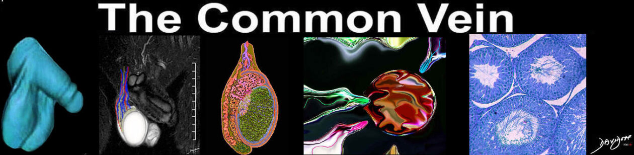
Vascular Diseases of the Testis
Authors Javier Gonzálezl Gaetano Ciancio
The arterial supply to each of the testis most often arises from the anterolateral surface of the aorta at L2-L3, just below the renal artery. Sometimes they may originate directly from the renal artery.
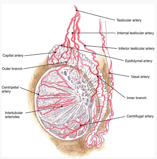
it divides into two main branches: (i) an outer branch, the internal testicular artery, and (ii) an inner branch, the inferior testicular artery. Surrounded by the pampiniform plexus , the tortuosity of the testicular arteries increases as they approach to the testicle. This configuration may be considered a heat-exchange system to cool the arterial blood at this level.
The inferior testicular artery runs between the posterior testis surface and the epididymal body, branching and entering through the posterior border of the testis at several different points. In contrast, a number of branches from the internal testicular artery pass through the tunica albuginea to enter the tunica vasculosa. Taken together these vessels form an aggregation which appears as a true vascular hilum in the posterior testicular aspect. Considerable variation is found in the branching and distribution of these vessels as they enter the testicular parenchyma, but they commonly ramify at the level of the tunica vasculosa to get inside the parenchyma as the coiled centripetal arteries. The centripetal arteries run toward the rete testis and, in turn, put out branches that completely reverse the course and return also as centrifugal arteries. Both sets of arteries divide further inside the parenchyma and end as intertubular arterioles (Cordier et al. 1938; Kormano 1967; Kormano and Suoranta 1971; McMinn 1995). The capillaries that arise from the arterioles enter the interstitial tissue and are separated from the germinal and supporting cells by a basement membrane that constitutes the “blood-testis barrier ” (Ploen and Setchell 1992).
The rete testis is therefore supplied by small vessels from the testicular mediastinum and from the small centripetal branches of the internal testicular artery. The tunica albuginea has its own set of capillary networks that are not related to the proper testis vascularization. Conversely, they arise independently from the branches of the testicular artery, from the vessels supplying the rete, and from the branches of the epididymal artery. The epididymal artery branches directly from the testicular artery at a level proximal to the epididymis. It supplies the head and the body of the epididymis through one or more capital arteries. The tail is vascularized by a complex network involving the epididymal, vasal, and testicular arteries, with supplementation from the cremasteric artery . This system provides an extensive anastomotic loop among these vessels (Cooper 1830; Hill 1909).
The artery of the vas deferens (i.e., deferential artery ) arises as a branch from the inferior vesical artery giving several branches to supply the vas deferens along its course and terminates with several capsular branches close to the mediastinum testis. Some of the vasal branches anastomose freely (i.e., following an irregular pattern) with branches of the testicular artery along the mediastinum and close to the lower end of the testis to form an epididymal-deferential loop (Harrison 1949; Yalçin et al. 2005).
The cremasteric artery arises from the inferior epigastric artery close to the deep inguinal ring to enter the inguinal canal supplying the cremasteric contents. It runs outside the internal spermatic fascia and so supplies an extremely limited amount of blood to the structures within. However, some terminal branches may reach the lower pole of the testis and anastomose with the epididymal-deferential loop , with a terminal branch of the testicular artery (80 %) or with branches of the epididymal artery (20 %) (Harrison 1949).
Vascular Testicular Areas
According to the amount of vascular supply, a number of different vascular areas can be distinguished in the testes: (i) rich vascular areas (upper polar, mediastinum testis, and posterolateral segments), (ii) moderate vascular areas (middle third of the lateral surfaces), and (iii) poor vascular areas (anterior border and anterolateral surfaces) (Harrison and Barclay 1948; Fig. 2).

Ashley Davidoff MD 133750
 Normal Testes Normal Testes |
| 47002c01 testis testes normal size blood flow normal anatomy USscan Davidoff MD |

Ashley Davidoff MD 133381

Ashley Davidoff MD 133379

47 M normal testes
Ashley Davidoff MD 133492

47 M normal testes
Ashley Davidoff MD
133490

Ashley Davidoff MD 133521

Ashley Davidoff MD 133525